Welcome back!
This week was our last week of testing our soil microbe, and we are fairly confident at this point that we have determined what our soil microbe is based on our previous tests. We believe our soil microbe to be Pseudomonas aeruginosa, an opportunistic pathogen that can infect humans and animals, but typically only infects immunocompromised individuals.
This week, we did an antibiotic test to try to determine our soil microbe. We used an antibiotic test medium and three controls, Staphylococcus aureus, Pseudomonas aeruginosa, and Escherichia coli plus our unknown (or somewhat unknown) soil microbe. We streaked the antibiotic plates with each microbe and used four antibiotics for each plate: carbenicillin at 100 micrograms, tetracycline at 30 micrograms, azithromycin at 15 micrograms, and erythromycin at 15 micrograms. The plates were left to incubate at 37 degrees Celsius for 24-48 hours. Unfortunately, our results for our plates were inconclusive and we did not get any data, and could not repeat the experiment due to a lack of supplies. However, had we had data, we would have expected to see sensitivity for our soil microbe, if it is P. aeruginosa, to the carbenicillin and no other antibiotics. P. aeruginosa has developed strong resistance to many antibiotics, making it difficult to treat and more likely to continue to develop resistance to other antibiotics as well. For E. coli, we would have expected to see sensitivity to carbenicillin, azithromycin, and erithromycin and resistance to tetracycline. The S. aureus control would have shown up as resistant to carbenicillin, which makes sense due to its Gram-negative nature; it would have also shown resistance to tetracycline, and sensitivity to azithromycin and erythromycin.
The last experiment that could be done to attempt to solidify our microbe would be to determine its ability or inability to liquefy gelatin; Pseudomonas strains of bacteria typically liquefy gelatin, so that would be one way to easily verify what we almost certainly already know, but to further determine the specific species of our microbe, we could determine its efficiency at growing at 37 degrees C; according to the dichotomous key, P. aeruginosa grows readily at 37-42 degrees C, and another similar strain, P. fluorescens, does not grow or grows poorly at 37 degrees C. By testing this, we could rule out one or the other strain depending on its ability to grow at this temperature.
I hope you've enjoyed following along as we have experimented these past few months and deduced what we think our soil microbe is!
Patricia
Monday, May 4, 2015
Tuesday, March 31, 2015
Week 6: Endospore Test Results
Welcome back to Week 6 of identifying our Soil Microbe!
Last week, Katie thought she may have been able to determine our soil microbe - this week, I will use the tests we did in last week's lab to determine if Katie was right, or if we need to keep testing to find our soil microbe.
This week in lab, we used two different methods to determine if our soil microbe contained endospores or not. Endospores are structures found inside the cell of some bacteria, usually Gram-positive Bacillus and Clostridium (Bauman 2014), that make cells more durable and resistant to death in unfavorable conditions as well as the potential to increase pathogenicity. Microbes have evolved to form endospores as a way to become more resistant to different methods of killing these pathogens and harmful microbiota and to increase their virulence to spread more rapidly.
For our lab, we used the Schaeffer-Fulton method of endospore staining to try and determine if and where in the cell our unknown soil microbe's endospore was located. To do this, we prepared 3 smears: our unknown microbe, a positive, endospore-forming control (Bacillus megaterium), and a negative, non-endospore-forming control (Escherichia coli). We heat fixed our smears, stained them with malachite green and then safranin, and observed them under the oil immersion objective lens of our microscope. When a microbe contains an endospore, it stains green while the rest of the cell stains the safranin, or pink, color. If there is not an endospore, the green will not be present, and the cells will all appear pink. However, the results of this staining method are frequently either inconclusive, or not strong enough to make a determination based on this one technique. Our results were completely inconclusive, and we were not even able to attain sufficient images of our microbe's cells to include as an example.
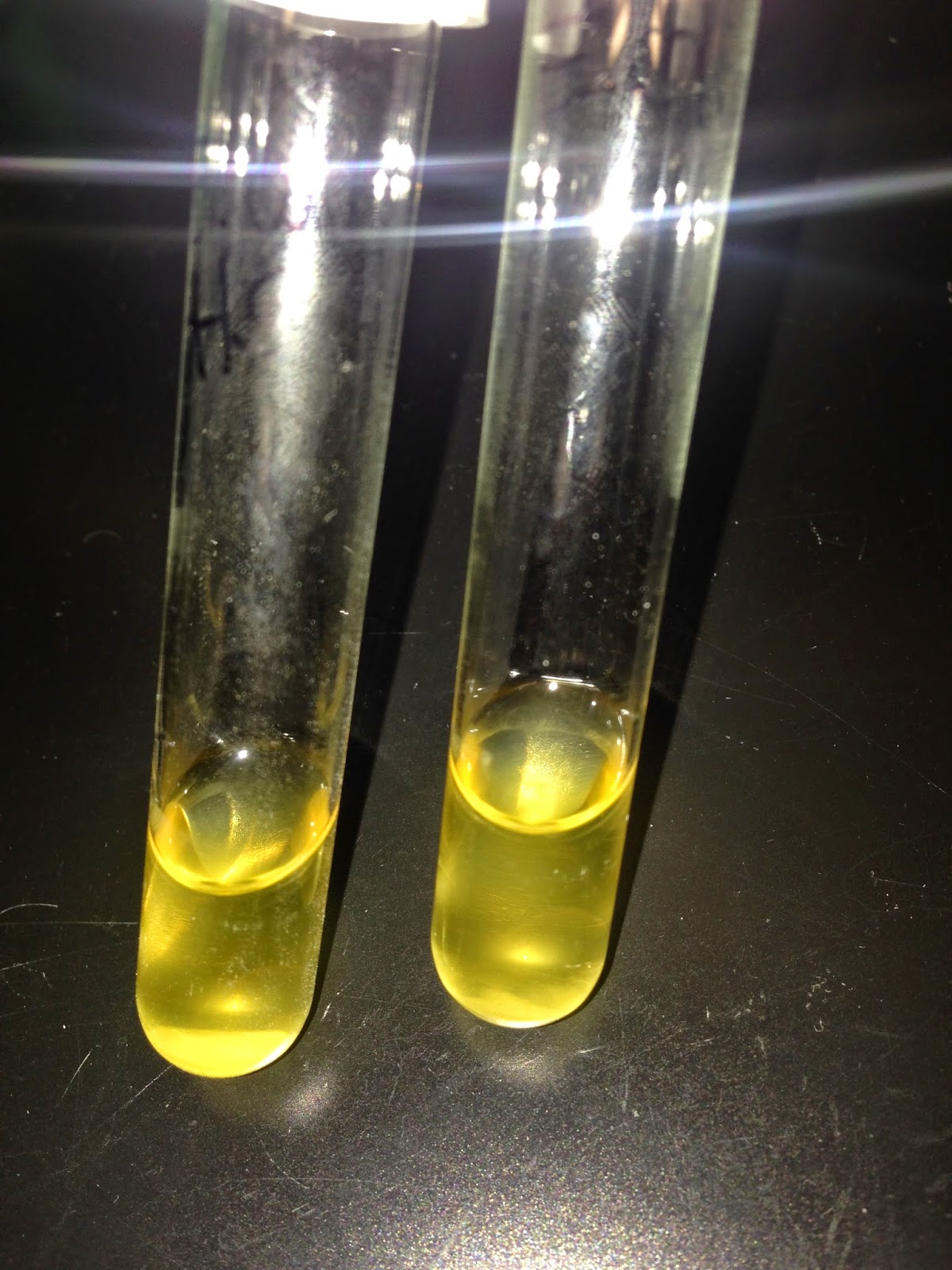
Because of the inconclusiveness of this staining procedure, we also used a heat shocking technique to determine if endospores form. Due to the high resistance of endospores, many microbes that contain them can be more resistant to higher temperatures. This means that, even after being put under high temperatures that would kill most normal microbes, endospore-forming microbes will still grow under these conditions. To determine if our microbe did, in fact, contain an endospore, we inoculated tubes containing TSB, a liquid growth medium for microbes, with each of our unknown soil microbe, our same positive control, and our negative control. We inoculated 2 tubes with each microbe, and heat shocked one tube of each microbe in an 80°C water bath for 10 minutes. After 4 days I came back to the lab to see if our microbe had grown under heat shock or not. Our microbe, as shown in the pictures below, grew in both the heat shocked and the regular tube, indicating that it is endospore-forming. The negative control, as you can see, is very clear and not cloudy at all in the heat shocked tube, but has bacteria growing in the tube incubated at room temperature. The B. megaterium, which is what we compared our unknown soil microbe against, also has growth in both tubes, therefore confirming that the similarity of our soil microbe to the positive control is due to our microbe being endospore-forming.
Using the dichotomous key from before, we can conclude that as Katie said, we more than likely have Pseudomonas aeuroginosa.
See you next week!
Patricia
Last week, Katie thought she may have been able to determine our soil microbe - this week, I will use the tests we did in last week's lab to determine if Katie was right, or if we need to keep testing to find our soil microbe.
This week in lab, we used two different methods to determine if our soil microbe contained endospores or not. Endospores are structures found inside the cell of some bacteria, usually Gram-positive Bacillus and Clostridium (Bauman 2014), that make cells more durable and resistant to death in unfavorable conditions as well as the potential to increase pathogenicity. Microbes have evolved to form endospores as a way to become more resistant to different methods of killing these pathogens and harmful microbiota and to increase their virulence to spread more rapidly.
For our lab, we used the Schaeffer-Fulton method of endospore staining to try and determine if and where in the cell our unknown soil microbe's endospore was located. To do this, we prepared 3 smears: our unknown microbe, a positive, endospore-forming control (Bacillus megaterium), and a negative, non-endospore-forming control (Escherichia coli). We heat fixed our smears, stained them with malachite green and then safranin, and observed them under the oil immersion objective lens of our microscope. When a microbe contains an endospore, it stains green while the rest of the cell stains the safranin, or pink, color. If there is not an endospore, the green will not be present, and the cells will all appear pink. However, the results of this staining method are frequently either inconclusive, or not strong enough to make a determination based on this one technique. Our results were completely inconclusive, and we were not even able to attain sufficient images of our microbe's cells to include as an example.
Because of the inconclusiveness of this staining procedure, we also used a heat shocking technique to determine if endospores form. Due to the high resistance of endospores, many microbes that contain them can be more resistant to higher temperatures. This means that, even after being put under high temperatures that would kill most normal microbes, endospore-forming microbes will still grow under these conditions. To determine if our microbe did, in fact, contain an endospore, we inoculated tubes containing TSB, a liquid growth medium for microbes, with each of our unknown soil microbe, our same positive control, and our negative control. We inoculated 2 tubes with each microbe, and heat shocked one tube of each microbe in an 80°C water bath for 10 minutes. After 4 days I came back to the lab to see if our microbe had grown under heat shock or not. Our microbe, as shown in the pictures below, grew in both the heat shocked and the regular tube, indicating that it is endospore-forming. The negative control, as you can see, is very clear and not cloudy at all in the heat shocked tube, but has bacteria growing in the tube incubated at room temperature. The B. megaterium, which is what we compared our unknown soil microbe against, also has growth in both tubes, therefore confirming that the similarity of our soil microbe to the positive control is due to our microbe being endospore-forming.
 |
| Our unknown soil microbe. Heat shocked culture is on the left. |
 |
| Our negative control, E. coli. Heat shocked culture is on the left. |
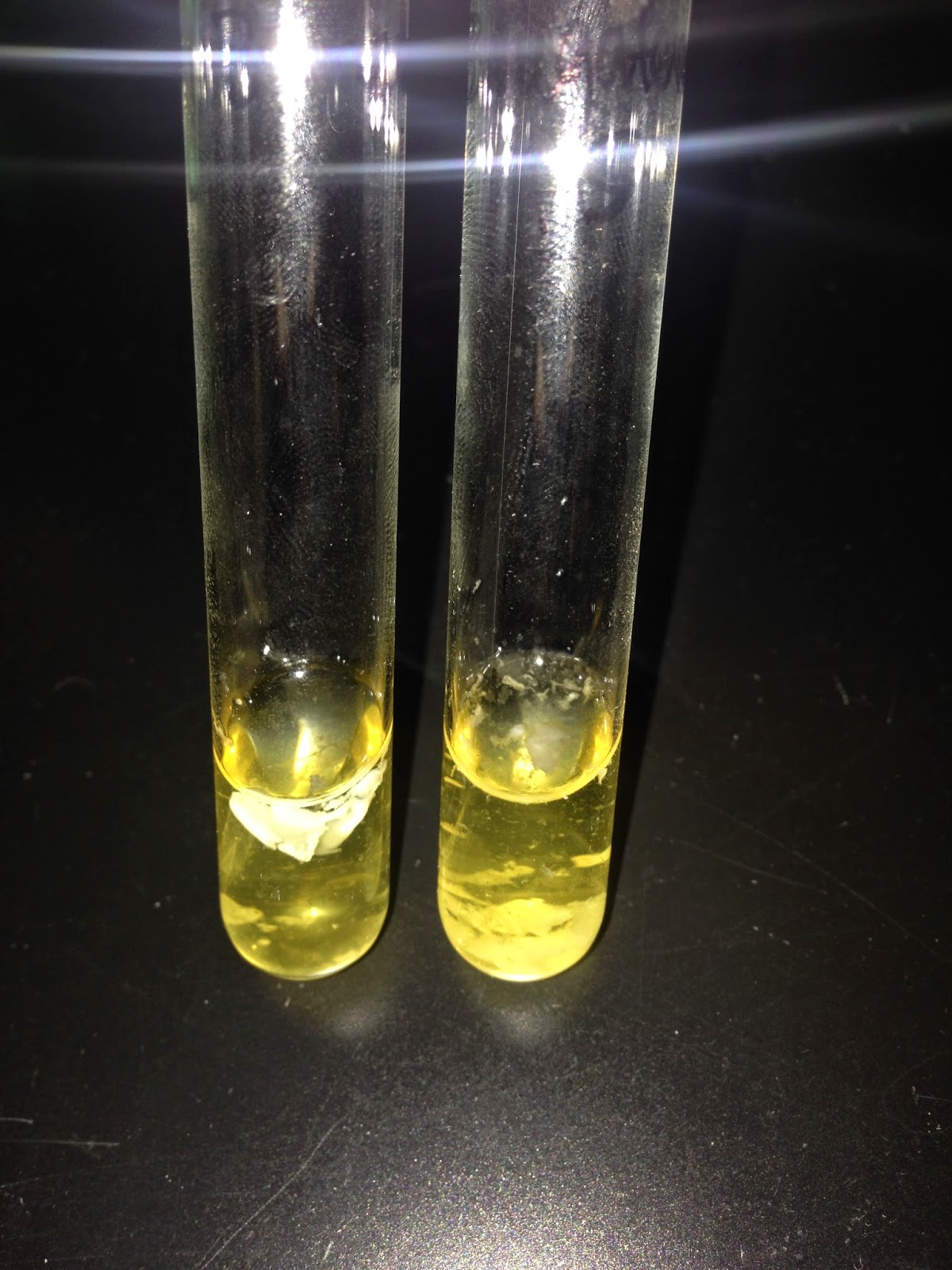 | |
| Our positive control, B. megaterium. Heat shocked culture is on the right. |
See you next week!
Patricia
Tuesday, March 10, 2015
Week 4: Acid-Fast Staining
Welcome back!
This week in lab, we worked on determining if our mysterious soil microbe was acid-fast or not. Acid-fast staining is a technique used to test for waxy cell walls in bacteria - it is similar to gram-staining because it involves using dyes to stain the cells, but the dyes are different and have different affects on the cells. If a bacteria in acid-fast staining stains a pink-ish color, the bacteria is said to be acid-fast, meaning it has a very waxy cell wall, and these are considered to be the micobacteria. If a bacteria in acid-fast staining stains a purple or blue color, the bacteria is said to be non-acid-fast, meaning it does not have a waxy cell wall.
We tested our soil microbe against some controls, which were all non-acid-fast. For our non-acid-fast control, Katie and I used B. megaterium, which is also a Gram-positive control that we used in last week's Gram-staining procedure. We expected our control to be a blue/purple color, and we compared the result of our control to our soil microbe and determined which type of acid-fast bacteria we assumed we had. An image of our soil microbe under the oil-immersion lens of the microscope is shown below:
As you can tell from this image, our microbe is clearly very non-acid-fast. The purple/dark blue color indicates to us that it is very non-acid-fast and thus has a thin waxy cell wall.
Using a dichotomous key, we can start to classify our microbe. I used a key provided by the professor, and since our microbe was Gram-negative, the next step on the dichotomous key is to determine if it is an anaerobic or aerobic, or facultatively anaerobic microbe. Since we have not done an experiment to determine this yet, we will have to wait to continue to classify our organism. Stay tuned until next week to see how we classify our microbe further!
Patricia
Tuesday, February 24, 2015
Week 2: Single Colony Purification
Welcome back!
This week, we checked back on our plates of soil microbes from last week and determined the number of colonies on the plate. We had a good bit of diversity on our plates, and found many different types of microbes. To determine the microbes per gram of soil on our plates, we loosely counted (visually) the number of colonies on each plate, and factoring in the dilutions. For the plates that were diluted at 10^-6 and 10^-7, no colonies were found, so these plates will be omitted from the experiment. For the other plates, our microbes per gram as soil were as follows:
10^-3 Plate: 65,000 microbes/g soil
10^-4 Plate: 140,000 microbes/g soil
10^-5 Plate: 7,000,000 microbes/g soil
Rose-Bengal agar plate (10^-4 dilution):10,000 microbes/g soil
We found it odd that our more diluted 10^-5 plate had more microbes/g soil than our 10^-4 plate, but we determined that this could be due to a larger number of fungi growing on the 10^-4 plate; we only observed one visible colony on the Rose-Bengal agar plate, but we did notice on the regular 10^-4 plate that what was growing on the Rose-Bengal plate appeared to also be growing, in one large colony, on the 10^-4 plate, which could have prevented our 10^-4 plate from growing more microbes than our more diluted 10^-5 plate.
There appeared to be a very decent, if not large, amount of biodiversity from our soil. There were smooth and rough surface appearances on microbes; circular, irregular, and rhizoid colony edges; and microbes with flat and convex elevations. This variety in surface appearance, colony edge, and elevation implies that every microbe that looks different from one another is probably a different microbe. Not only that, even some of the microbes that looked very similar can also be different from one another and is not easily determinable from the human eye's perspective. The large amount of biodiversity in this small sample of soil can show us that the biodiversity surrounding us is massive and we don't even realize it.
So, why does biodiversity matter? Biodiversity is defined as the variety of life on Earth. This variety leads us to make new discoveries, utilize the naturally occurring elements around us, and take advantage of all that the Earth can provide to us to help us survive. The biodiversity on Earth matters because humans are all dependent on the benefits we receive from our environment - the air we breathe, the foods we grow, the animals we raise for food, the naturally occurring elements that help us find cures to diseases and to live healthier lives. Specifically in soil, soil microbes are important in Nitrogen-fixation, which is necessary for the growth of many important organisms including many of the plants and vegetables we eat. Soil microbes can also help break down organic matter that would not naturally break down, helping us to recycle many nutrients that would normally be thrown away. All of these these reasons for biodiversity show us that the earth is a fascinating, diverse, and complex place, and without the study of biodiversity and things such as soil microbes, we would not be able to take advantage of all that is naturally occurring around us and make advances in studies, especially microbiology.
See you next week!
Subscribe to:
Posts (Atom)